
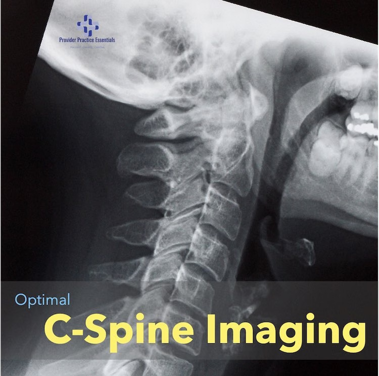
Imaging the cervical spine is crucial for diagnosing and managing various conditions, including trauma and degenerative disorders. Physicians often rely on several imaging modalities to achieve accurate diagnoses. This article explores the different imaging options, their strengths and weaknesses, and the potential for combining modalities to identify fractures. Additionally, we’ll delve into the reliability of AI in detecting cervical spine fractures.
Imaging Modalities
- X-rays: X-rays are the initial imaging choice for cervical spine evaluation. They provide a quick and cost-effective way to assess bony structures, including fractures, dislocations, and degenerative changes. However, they are limited in their ability to visualize soft tissues and may miss subtle injuries.
- Computed Tomography (CT): CT scans offer detailed images of both bone and soft tissues. They are highly sensitive in detecting fractures, especially in complex cases or when assessing spinal stability. CT is often used when a fracture is suspected but not visible on X-rays.
- Magnetic Resonance Imaging (MRI): MRI is unparalleled in assessing soft tissues like spinal cord, ligaments, and discs. It is invaluable for evaluating spinal cord injuries, disc herniations, and ligamentous damage. However, it may not be the best choice for initial trauma assessment due to time constraints and limited bony detail.

Strengths and Weaknesses
- X-rays: Strengths include rapid imaging and minimal radiation exposure. Weaknesses include limited soft tissue visualization and reduced sensitivity for subtle injuries.
- CT: Strengths include high-resolution images of bones and the ability to detect fractures accurately. Weaknesses include higher radiation exposure and limited soft tissue contrast.
- MRI: Strengths include excellent soft tissue contrast and absence of ionizing radiation. Weaknesses include longer acquisition times, patient claustrophobia, and contraindications for certain patients (e.g., those with metallic implants).
Combining Modalities for better Cervical Spine Imaging
Combining imaging modalities can enhance diagnostic accuracy, especially in complex cases. For example, a combination of X-rays and CT scans can provide a comprehensive assessment of both bone and soft tissue structures. This approach is valuable when assessing spinal stability and planning surgical interventions.
AI in Fracture Detection
Artificial intelligence (AI) has made significant strides in medical imaging, including fracture detection. AI algorithms can assist physicians by quickly and accurately identifying fractures on imaging studies. These AI systems can analyze large datasets, detect subtle fractures, and reduce the risk of oversight. However, it’s essential to remember that AI is a supplementary tool, and human expertise remains indispensable for clinical decision-making.
Academic Centers Leading the Way in Cervical Spine Imaging
Major academic medical centers play a pivotal role in advancing the field of cervical spine imaging. These institutions are at the forefront of research, innovation, and clinical practice, continually pushing the boundaries of diagnostic capabilities. Here are some notable initiatives and trends:
Advanced Imaging Protocols
Academic centers are known for developing and implementing cutting-edge imaging protocols. They are often early adopters of new technologies, such as ultra-high-resolution CT scanners and advanced MRI sequences. These innovations aim to improve image quality, reduce radiation exposure, and enhance diagnostic accuracy.
Multidisciplinary Collaboration
Collaboration between radiologists, neurosurgeons, orthopedic surgeons, and other specialists is a hallmark of academic centers. This interdisciplinary approach ensures that the full spectrum of cervical spine conditions is considered during diagnosis and treatment planning.
Research in AI and Machine Learning
Many academic centers are actively involved in research related to artificial intelligence and machine learning in medical imaging. They are developing and testing AI algorithms specifically tailored to cervical spine imaging. These algorithms have the potential to streamline the diagnostic process and increase the detection rate of fractures, especially in complex cases.
Clinical Trials
Academic institutions often lead clinical trials to evaluate the effectiveness of new imaging techniques or treatment approaches for cervical spine disorders. These trials contribute valuable data to the medical community and help establish evidence-based guidelines.
Education and Training
Academic centers are hubs for medical education and training. They train the next generation of physicians, radiologists, and technicians in the nuances of cervical spine imaging. Moreover, they provide continuous education opportunities for healthcare professionals to stay updated with the latest advancements.
Patient-Centered Care
Academic centers prioritize patient-centered care, focusing on personalized treatment plans tailored to each patient’s unique needs. This approach often involves a thorough assessment using a combination of imaging modalities to ensure the most accurate diagnosis and optimal treatment outcomes.
Telemedicine and Remote Consultations
In response to the global shift towards telemedicine, academic centers have embraced remote consultations and virtual imaging reviews. This enables expert consultation for complex cases, even across geographical boundaries, ensuring that patients receive the best possible care.
Major academic medical centers are driving progress in cervical spine imaging through their commitment to research, multidisciplinary collaboration, and the integration of innovative technologies. They serve as vital hubs for education, clinical trials, and the development of AI tools, ultimately benefiting both healthcare professionals and patients alike. By staying at the forefront of medical imaging, these institutions continue to shape the future of cervical spine diagnosis and treatment.
Conclusion
Selecting the optimal imaging method for the cervical spine depends on the clinical scenario and the information required. X-rays are useful for initial assessment, CT scans excel at detecting fractures, and MRI provides detailed soft tissue evaluation. Combining modalities can enhance diagnostic accuracy, while AI offers promising support in fracture detection. Physicians must consider patient-specific factors and clinical judgment when choosing the most appropriate imaging approach for cervical spine evaluation.


